
Ever wondered how an X-ray machine actually “sees”? While we often focus on the image sensor, the process actually begins with a critical component called the scintillator.
The Physics of Conversion
At its core, a scintillator is a specialized material that exhibits luminescence when excited by ionizing radiation. When X-ray photons strike the scintillator, they are absorbed and re-emitted as visible light. This light can then be captured by high-performance CMOS or CCD image sensors to create the digital images we rely on.
Material Excellence
The choice of material is vital for determining the quality, speed, and sensitivity of the imaging system. Common high-performance materials include:
- CsI(Tl) (Cesium Iodide): Known for its excellent light yield and high resolution.
- YAG(Ce) & LYSO: Preferred for high-speed applications due to their fast decay times.
- GOS (Gadolinium Oxysulfide): A robust choice for various industrial and medical X-ray detectors.
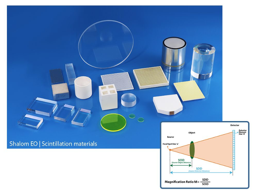
System Precision and Resolution
Beyond the material itself, achieving high-resolution imaging requires a deep understanding of system geometry. Factors such as magnification, pixel size, and the thickness of the scintillator layer play a decisive role in minimizing blur and maximizing clarity.
Why It Matters
For engineers and system integrators in the medical, dental, and industrial inspection sectors, selecting the right scintillator-sensor combination is the key to developing next-generation imaging solutions.